Comparto unos apuntes de la sesión clínica sobre introducción a la ecografía en Atención Primaria que espero que motive a mis compañeras de equipo para incorporar esta técnica a su práctica clínica.
Objetivo de la sesión: motivar.
Quién la imparte? Un novato con ganas de aprender. Un médico de familia que empezó hace año y medio a hacer ecografías. Os cuento qué he aprendido:
- Fundamentos.
¿Qué es la ecografía? Tecnología aplicada al diagnóstico (anamnesis, exploración física, ecoscopia)
¿Para qué sirve?. Para ver un poco más. Complementa la exploración, afina el dx..
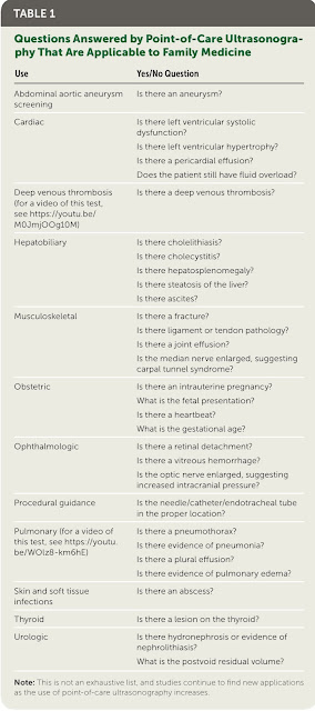
¿Qué es lo más difícil?: la indicación. Diferenciar las exploraciones que precisa el médico para aprender de las que necesita el paciente en su proceso. Hacernos siempre la pregunta: ¿Qué quiero descartar haciendo esta ecografía? (Tabla 1)
Ventajas: accesibilidad, rapidez, potencia diagnóstica
Desventajas: tiempo invertido en formación. tiempo de ejecución. Falsos positivos y negativos. Ley de cuidados inversos, ml uso de tiempo y recursos (exceso de peticiones de ecografías regladas, o cadenas diagnósticas). Sobrediagnóstico, sobretratamiento, iatrogenia.
- ¿Cómo empezar? plan personal de formación.
Cursos. SERMAS, Semfyc, Semergen…
Artículos.
Libros.
Vídeos.
RRSS.
Compaginar teoría y práctica.
Tener paciencia: hay que pasar de la ceguera ecográfica a la visión ecográfica.
- ¿Qué necesitas?
Un ecógrafo.
Conocimiento básico de técnica.
Repaso anatómico.
Conocimiento de ecoscopia normal.
Conocimiento de ecoscopia patológica.
Practicar, practicar, practicar.
- Objetivo:
hacer ecoscopias breves (2-5 minutos) en consulta a demanda.
Adecuar número diario o semanal según presión asistencial y circunstancias profesionales.
También se pueden programar y reservar más tiempo.
Llegar a ser ecoscopista no ecografista ni radiólogo (igual que aprendiste a leer ekg sin ser cardiólogo)
- ¿Cómo conseguirlo? Perseverancia. Subir la montaña, un paso de teoría otro de práctica. Empezar por el principio. Plan de acción: Reservar 10-20 minutos semanales para programar 1-2 exploraciones todas las semanas. Registrar siempre: en protocolo ecografía APMadrid a ser posible.
Hacer fotos y vídeos y grabarlos en el ecógrafo o en el móvil.
Resolver dudas, preguntando a compañeros, esperando la ecografía reglada.
Acostumbrarse a ver todas las ecografías que te lleguen informadas o hagan a tus pacientes en el hospital (no desperdiciar la docencia indirecta del servicio de radiología). Si ves algo interesante compártelo.
Un libro:
https://es.scribd.com/document/579697057/Ecografi-a-a-pie-de-cama-Nilam-J-2%C2%AA-Edicio-n-2020-1
https://m.youtube.com/watch?si=65Z-mWAAUEknvcxW&v=fyakQ4LM5sM&feature=youtu.be&cbrd=1
https://www.ultrasoundtraining.com.au/resources/coaching-corner/
General Medicine, Internal Medicine & Primary Care Ultrasound
Broome Docs – Rural Generalist Doctors Education by Casey Parker.
Gastric Ultrasound – everything you need to know.
IMBUS Program – Abbott Northwestern Hospital – Medical Education Department.
NephroPOCUS.com – Abhilash Koratala (ssociate Professor of Medicine and currently the Director of Clinical Imaging for Nephrology at the Medical College of Wisconsin) created this website to advocate for point-of-care ultrasonography (POCUS)-enhanced physical examination and serve as a comprehensive resource for anybody interested in POCUS, particularly pertaining to nephrology practice.
POCUS101 – Point of Care Ultrasound Made EASY – lead by Vi Dinh.
POCUS Med Ed – tutorials, cases, research.
Sonointernest.com – internal medicine POCUS by Michael Wagner.
Ultrasound G.E.L. (Gathering Evidence from the Literature) – a podcast and website for learning about recent literature in the field of point-of-care ultrasound.
Ultrasound Image/Case Sites
Core Ultrasound – ultrasound of the week.
EM Cases: POCUS – real cases from the ED.
Everyday Ultrasound – A crowdsourced page – images and videos of cool cases with quick #POCUS pearls.
Grepmed – Grepmed is a crowdsourced medical image site which has a huge POCUS image library. If you’re looking for sonographic pathology or putting together a presentation, this is a great website to visit.
Life in the Fast Lane – LITFL 100 Ultrasound quiz.
SonoDoc (A Bedside Ultrasound Game) – A fun, interactive, patient-centered and case-based game providing instruction on the indications, technique, and interpretation of various POCUS applications, from leading POCUS expert, Dr. Laleh Gharahbaghian and team at Stanford Med. CME available for those eligible too!
The Pocus Atlas – content is freely available to share and use for global point-of-care ultrasound education.
UltrasoundBox – An interactive Ultrasound Education Platform.
Ultrasoundcases.info – loads of cases free offered to you by SonoSkills and FUJIFILM Healthcare Europe.
Ultrasound imaging – exactly what it says.
Ultrasounodpaedia – Your portal to a world of ultrasound education and training
RECURSOS EN TWITTER
Mi lista de ecografía:
https://x.com/i/lists/1590329722050940928
Yale Tung
Joaquín Martínez
https://x.com/y_interna/status/1760026640573157468?s=61&t=i-aqOCWtRO2zssEdvpTdCA
https://x.com/y_interna/status/1759669914967687392?s=61&t=i-aqOCWtRO2zssEdvpTdCA
https://x.com/y_interna/status/1757469199494991913?s=61&t=i-aqOCWtRO2zssEdvpTdCA
https://x.com/jmmr83/status/1785980616992809100?s=61&t=i-aqOCWtRO2zssEdvpTdCA
ecografía AP
https://www.coreultrasound.com/5ms/
https://www.aafp.org/dam/brand/aafp/pubs/afp/issues/2018/0815/p200.pdf
Abdomen:
https://www.youtube.com/watch?v=uob37av7nSQ
https://www.youtube.com/watch?v=lsDVzWu_UdQ
Introduction to Primary Care Ultrasound.
I'm sharing some notes from the clinical session on an introduction to ultrasound in Primary Care that I hope will motivate my team members to incorporate this technique into their clinical practice.
Session objective: to motivate.
Who delivers it? A novice eager to learn. A family doctor who started doing ultrasounds a year and a half ago. Let me tell you what I've learned:
1. Fundamentals.
What is an ultrasound? Technology applied to diagnosis (history taking, physical examination, echoscopy).
What is it used for? To see a bit more. It complements the examination, refines the diagnosis.
What is the most challenging part? The indication. Differentiating the examinations that the doctor needs to learn from those that the patient needs in their process. Always ask ourselves: What do I want to rule out by doing this ultrasound? (Table 1)
Advantages: accessibility, speed, diagnostic power.
Disadvantages: time invested in training, execution time, false positives and negatives, inverse care law, misuse of time and resources (excessive requests for scheduled ultrasounds, or diagnostic chains), overdiagnosis, overtreatment, iatrogenesis.
1. How to start? Personal training plan.
Courses. SERMAS, Semfyc, Semergen...
Articles.
Books.
Videos.
Social networks.
Combine theory and practice.
Be patient: you have to move from echographic blindness to echographic vision.
1. What do you need?
An ultrasound machine.
Basic knowledge of technique.
Anatomical review.
Knowledge of normal echoscopy.
Knowledge of pathological echoscopy.
Practice, practice, practice.
1. Goal:
to perform brief echoscopies (2-5 minutes) in consultation on demand.
Adjust the number daily or weekly depending on healthcare pressure and professional circumstances.
You can also schedule and reserve more time.
Aim to become an echoscopist, not an ultrasonographer or radiologist (just as you learned to read EKGs without being a cardiologist).
1. How to achieve it? Perseverance. Climbing the mountain, one step of theory, one step of practice. Start from the beginning. Action plan: Reserve 10-20 minutes weekly to schedule 1-2 explorations every week. Always record: follow the APMadrid ultrasound protocol if possible.
Take photos and videos and save them on the ultrasound machine or mobile.
Resolve doubts by asking colleagues, waiting for the scheduled ultrasound.
Get used to seeing all the ultrasounds that are reported or performed on your patients in the hospital (do not waste the indirect teaching from the radiology department). If you see something interesting, share it.
A book:
https://es.scribd.com/document/579697057/Ecografi-a-a-pie-de-cama-Nilam-J-2%C2%AA-Edicio-n-2020-1
https://m.youtube.com/watch?si=65Z-mWAAUEknvcxW&v=fyakQ4LM5sM&feature=youtu.be&
初级保健超声波简介
机器翻译,如有错误,敬请原谅。
与耶鲁·唐、伊格纳西奥和拉法·阿隆索·罗卡一起。
我分享了一些关于初级保健超声引入的临床会议笔记,希望这能激励我的团队成员将这项技术纳入他们的临床实践。
会议目标:激励。
谁来主持?一位渴望学习的新手。一位家庭医生,一年半前开始做超声检查。让我告诉你我学到了什么:
1. 基础知识。
什么是超声?应用于诊断的技术(病史采集、体格检查、超声检查)。
它有什么用?能看到更多。它补充了检查,精细化了诊断。
最难的部分是什么?指示。区分医生为了学习而需要的检查和病人在其过程中需要的检查。我们始终要问自己:我做这个超声检查想排除什么?(表1)
优点:易于获取、速度快、诊断力强。
缺点:投入培训时间、执行时间、假阳性和假阴性、逆向关怀法则、时间和资源的误用(超声检查的过度请求或诊断链)、过度诊断、过度治疗、医源性损害。
1. 如何开始?个人培训计划。
课程。SERMAS, Semfyc, Semergen...
文章。
书籍。
视频。
社交网络。
结合理论与实践。
要有耐心:你必须从超声盲转变为超声视觉。
1. 你需要什么?
一台超声机。
基础技术知识。
解剖复习。
正常超声检查知识。
病理超声检查知识。
实践,实践,再实践。
1. 目标:
根据需求在咨询中进行简短的超声检查(2-5分钟)。
根据医疗压力和专业情况调整每日或每周的数量。
你也可以安排并预留更多时间。
目标是成为超声检查者,而不是超声技师或放射科医师(就像你学习读心电图而不是成为心脏病学家一样)。
1. 如何实现?坚持不懈。爬山,理论一步,实践一步。从头开始。行动计划:每周预留10-20分钟,每周安排1-2次探查。尽可能始终记录:遵循APMadrid超声协议。
拍照和视频,保存在超声机或手机上。
通过询问同事、等待预定的超声来解决疑问。
习惯于看到在医院对你的病人进行的所有报告或执行的超声(不要浪费放射科的间接教学)。如果你看到有趣的东西,分享它。
一本书:
https://es.scribd.com/document/579697057/Ecografi-a-a-pie-de-cama-Nilam-J-2%C2%AA-Edicio-n-2020-1


No hay comentarios:
Publicar un comentario
También puede comentar en TWITTER a la atención de @DoctorCasado
Gracias.
Nota: solo los miembros de este blog pueden publicar comentarios.